Iron Deficiency vs. Thalassemia Minor
------------------------------------------------------------------------
*Iron Deficiency* *Thalassemia Minor* *Combined*
*MCV* Low Very Low Very Low
*RDW* High Normal or High High
*Red Cell Count* Low Normal or High Normal or Low
*Marrow Iron* Absent Normal Absent
*Serum Iron* Low Normal Low
*TIBC* High Normal High
*Serum Ferritin* Low Normal Low
*Hemoglobin A2* Low High Normal
 Normal Values
 RBC count (varies with altitude):
Male: 4.7 to 6.1 million cells/mcL
Female: 4.2 to 5.4 million cells/mcL
WBC count: 4,500 to 10,000 cells/mcL
Hematocrit (varies with altitude):
Male: 40.7 to 50.3%
Female: 36.1 to 44.3%
Hemoglobin (varies with altitude):
Male: 13.8 to 17.2 gm/dL
Female: 12.1 to 15.1 gm/dL
RDW <14.5%
MCV: 80 to 95 femtoliter
MCH: 27 to 31 pg/cell
MCHC: 32 to 36 gm/dL

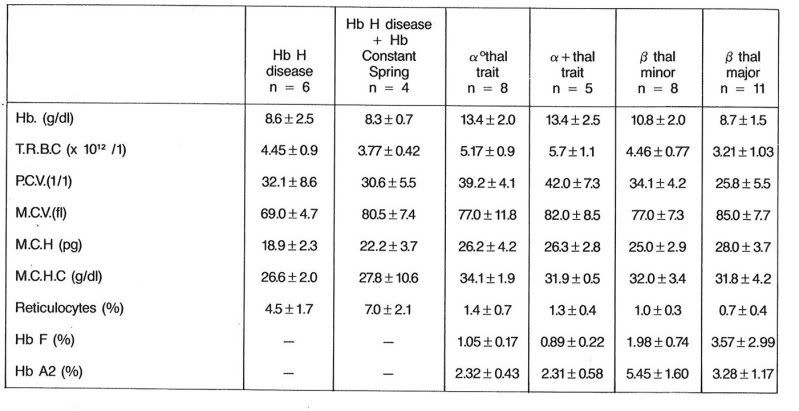
The reticulocyte count may assist in differentiating a hyporegenerative or arregenerative anemia from one due to RBC destruction (Irwin & Kirchner, 2001; Lesperance et al., 2002). An elevated reticulocyte count suggests premature release of immature RBCs into the circulation (Abshire, 2001) to replace losses from rapid destruction, as occurs in hemolytic disorders. Conversely, a low reticulocyte count in the face of anemia suggests decreased production or release of RBCs (Cohen, 1996), as may be found in bone marrow failure, iron deficiency, lead poisoning, and anemia of inflammation (Hermiston & Mentzer, 2002). Assessment of the reticulocyte count is particularly helpful in the diagnosis of anemia of prematurity, as it will be paradoxically decreased for the degree of anemia present. In addition to its diagnostic potential, the reticulocyte count may be used to guide response to anemia management, including nutritional support or exogenous erythropoietin administration (Abshire, 2001; Widness, 2000).